Results
Photographs of work carried out by Dr Singh
Case gallery
Filter results by treatment
Showing 11 cases.
 Before
 After
AfterCrowding corrected with clear aligners
Crowded and rotated upper front teeth brought into line with clear aligners, and the shade lifted afterwards to finish the result. Photographed in the same position and the same light before treatment and at completion.
 Before
 After
AfterUpper front teeth rebuilt with composite bonding
Short upper front teeth with worn, uneven edges rebuilt in composite, shaped tooth by tooth to even out the edge line and the proportions. Photographed before treatment and at completion.
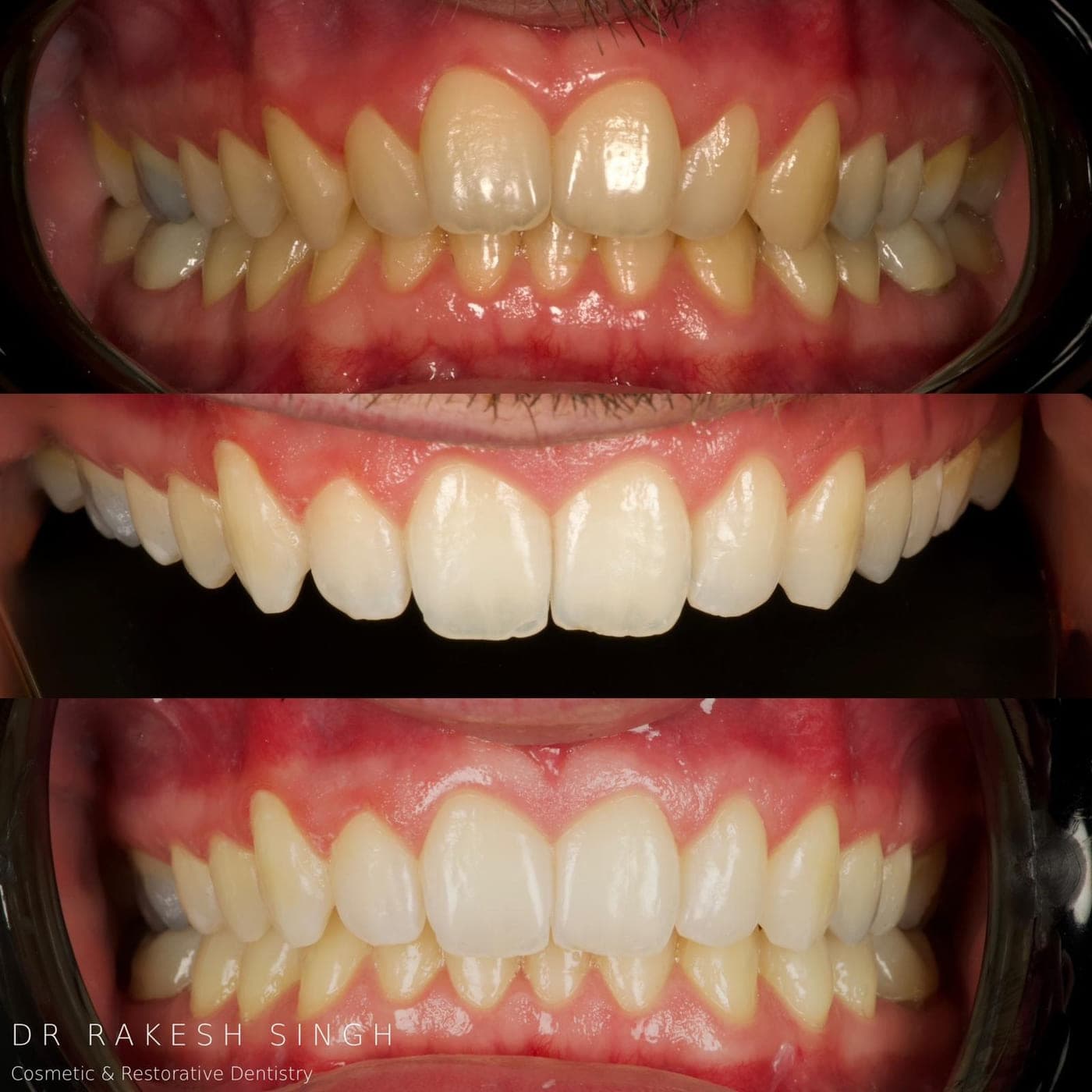
Anterior restoration, stage by stage
A three-stage clinical record of the upper and lower front teeth: the starting position, the restored upper teeth under isolation, and the completed result in the bite.
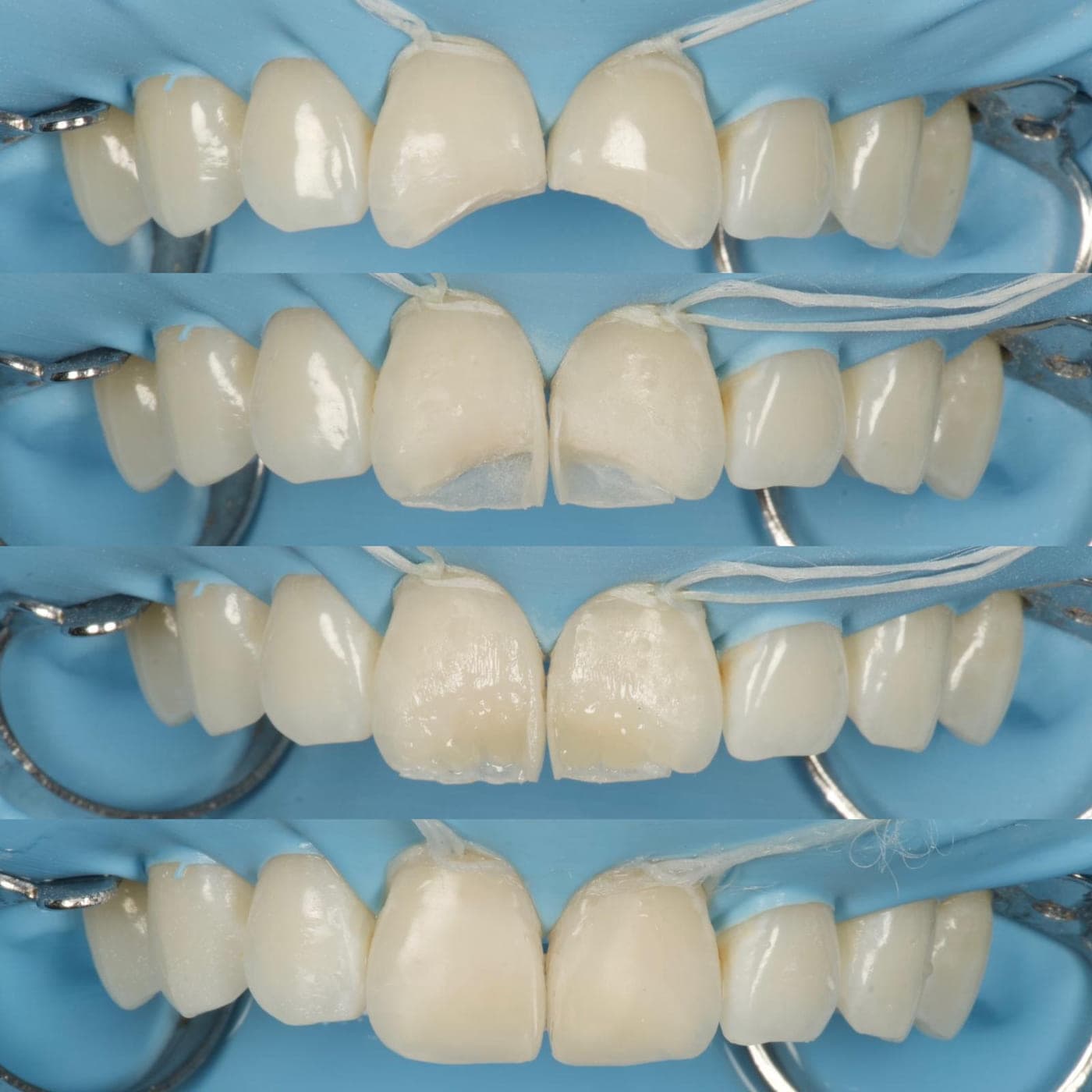
Composite build-up under isolation
A four-stage record of composite being placed on two upper central incisors under rubber dam, from the prepared surface through to the shaped and polished result.
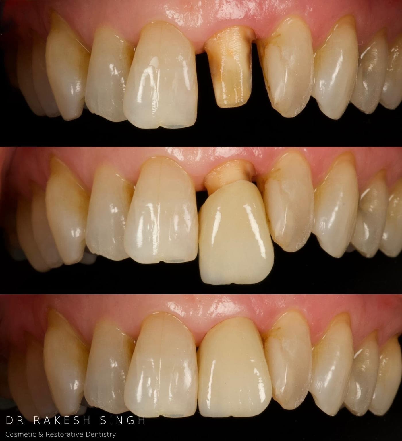
Single crown on a discoloured front tooth
A darkened upper front tooth prepared, then restored with a crown matched to the neighbouring teeth. The middle frame shows the crown at try-in.
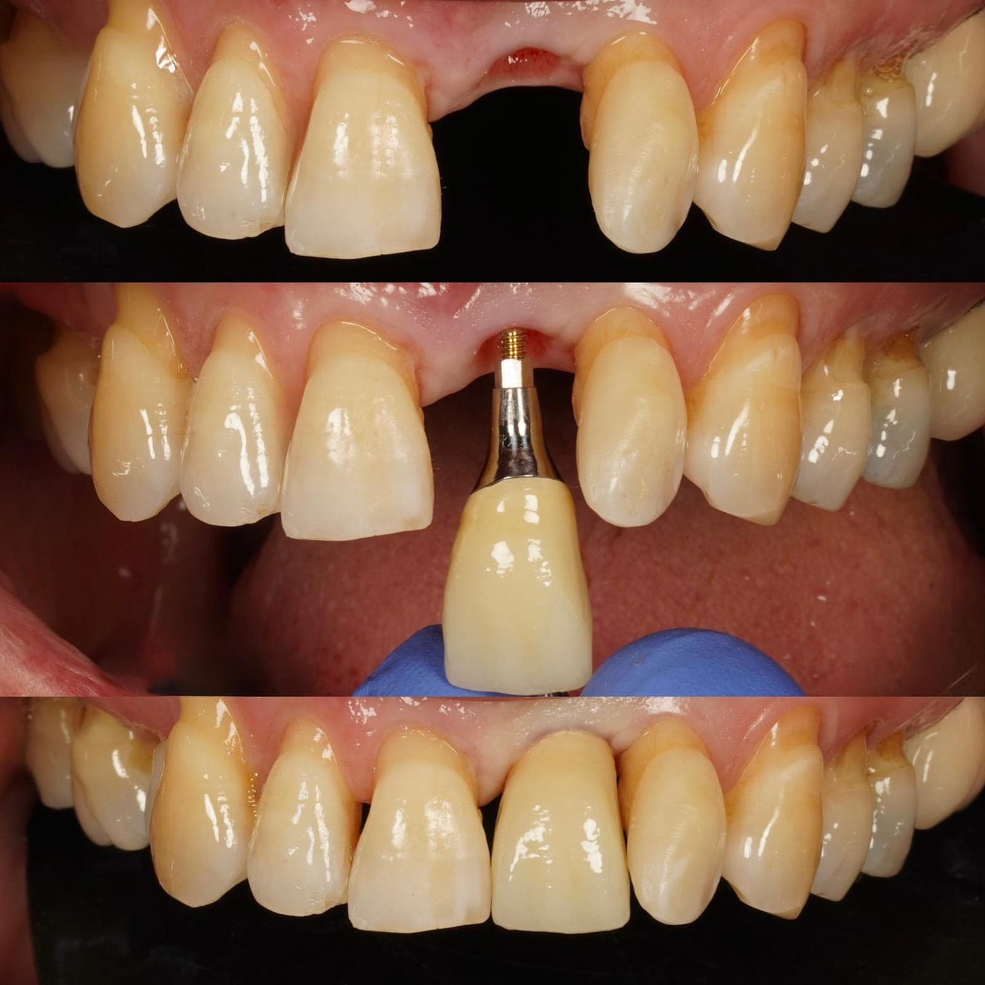
Implant crown replacing a single upper tooth
A missing upper front tooth replaced with an implant-supported crown. The teeth either side were not prepared. The middle frame shows the crown and abutment before fitting.
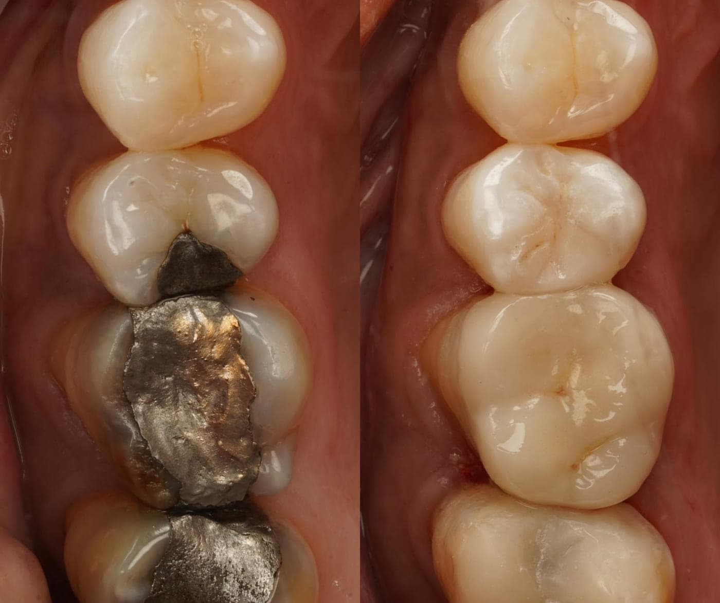
Metal fillings replaced with tooth-coloured restorations
Large, worn metal fillings in the back teeth replaced with tooth-coloured restorations, rebuilding the shape of each tooth and its contact with its neighbour.
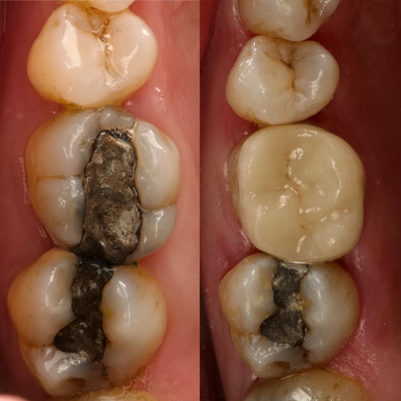
Ceramic onlay replacing a failing metal filling
A heavily filled molar restored with a ceramic onlay covering the biting surface, rather than a further direct filling.
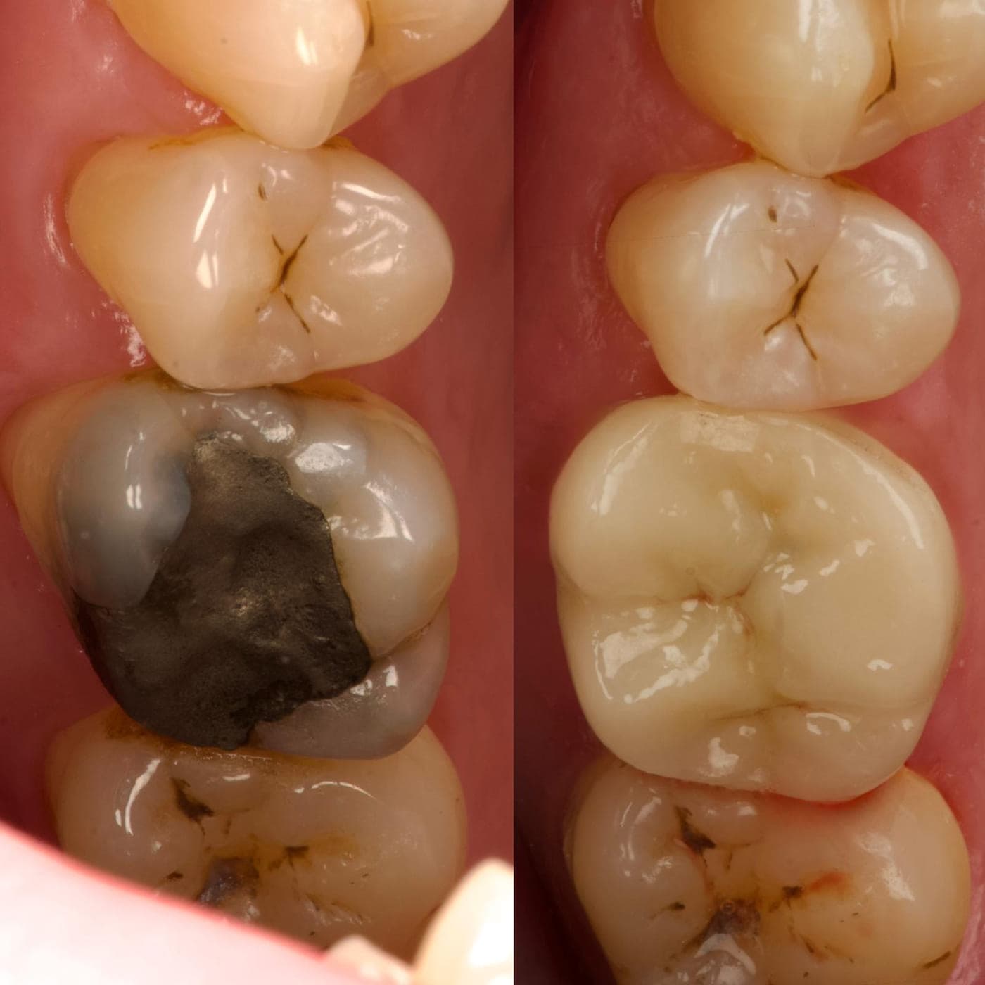
Rebuilding a broken-down molar
A molar with a large failing metal filling restored to its original contours with a tooth-coloured restoration.
 Annotated by the practice
Correcting the upper edge line
The upper edge line brought into a curve that follows the lower lip. Annotated by the practice: the red line marks the starting edge position, the green line the position after treatment.
 Annotated by the practice
Edge position, shown at magnification
The same change to the upper edge line shown with the practice’s own magnified detail of the upper right teeth.
Dental treatment is planned for the individual. Whether any treatment described here is appropriate for you can only be established at a clinical assessment, and the options discussed with you may differ from those set out on this page. Photographs show the outcome for one patient and are not a prediction of anyone else’s result.
Ask what is realistic for your teeth.
Photographs show what was possible for one person. What is possible for you is a question for an examination.